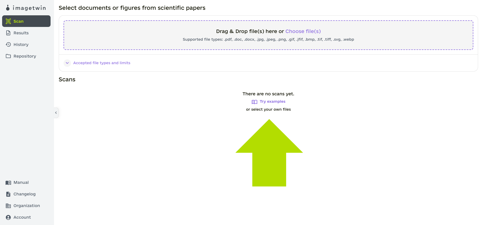
Drag and drop your file(s) into the dedicated area or click the area and choose a file through a file selection dialog.
Supported formats include PDF, DOCX, DOC, JPG, PNG, GIF, BMP, TIF, JFIF, SVG, and WEBP.
Step 3
After reviewing the selected files, click “Scan” at the bottom of the page. Scans are automatically saved for 90 days and can be accessed in the History tab (on the left side). If you prefer not to save a scan, simply uncheck the “Save scans for 90 days” option before proceeding. Saved scans are only accessible and visible to you (and/or your organisation).
Step 4
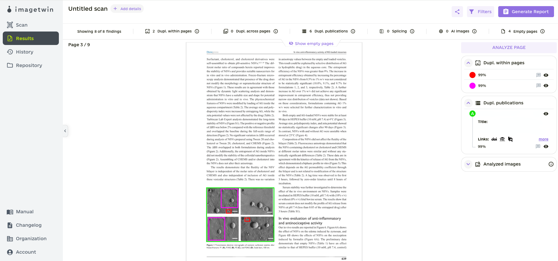
Congratulations! The scan has been successfully completed. Here is what the result page will look like:
Step 5
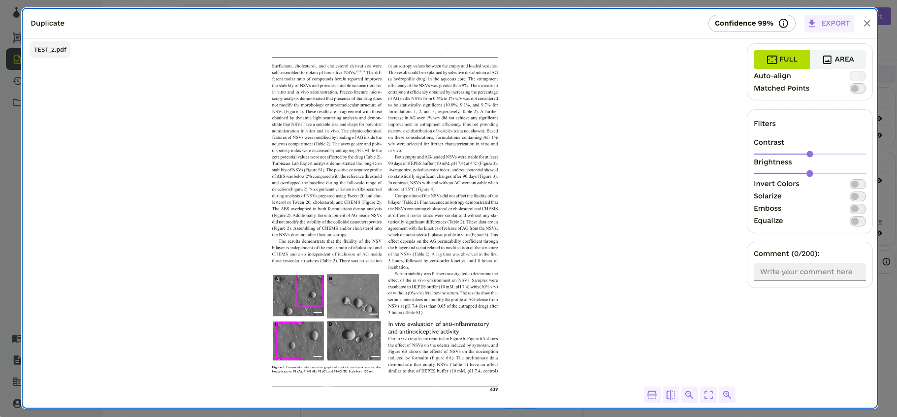
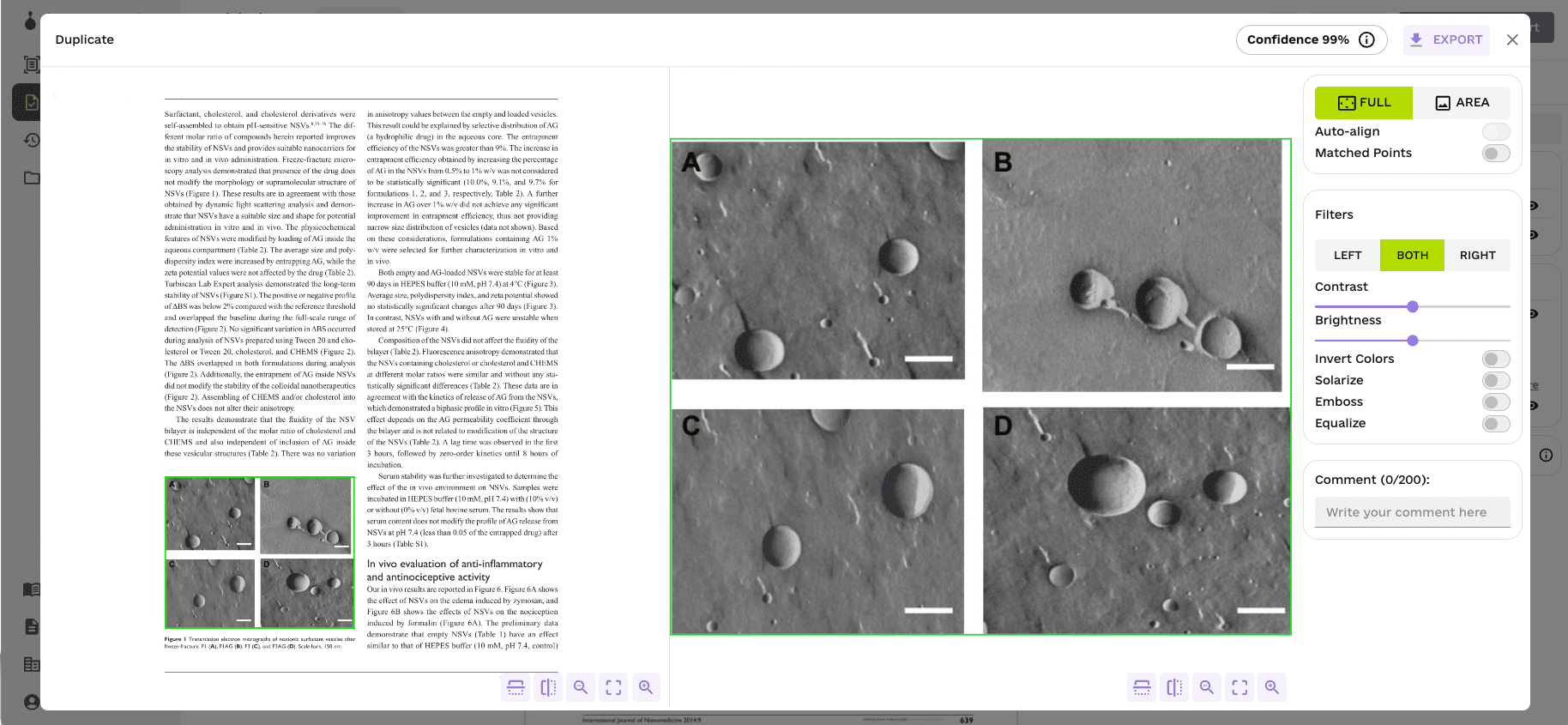
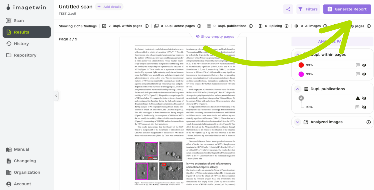
This is where you can review each page. The app will show you where it has detected duplicates within the paper you have uploaded or across other publications. By clicking on a finding, the detail view opens. The details view allows you to investigate the finding further and offers a forensic toolbox.
If the app has detected a duplicate within your publication, it will mark the area with a colored bounding box.
If the app has detected a duplicate across external publications, it will mark the area with a colored bounding box.
Other Features
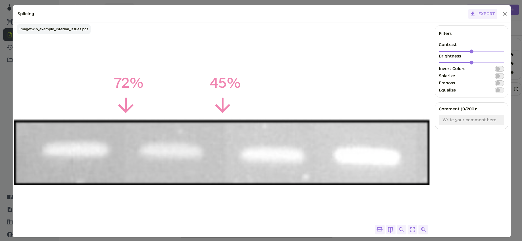
Splicing detection
The software detects vertical splicing in gel band images, characterized by sharp edge transitions. Based on our detection model’s confidence, it will display scores from 0% (low) to 100% (high confidence). This is what the detail view looks like for splicing:
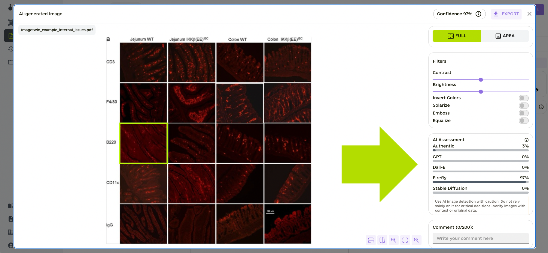
AI-Generated Image Detection
Each paper checked by Imagetwin will also be scanned using our AI detector for images to help users identify AI-generated visuals.
Each flagged image includes an “AI assessment” showing a distribution of which generator was most likely used. For example, you may see a score indicating a higher probability of generation by Firefly compared to DALL·E or Stable Diffusion. This gives users more context to evaluate flagged cases.
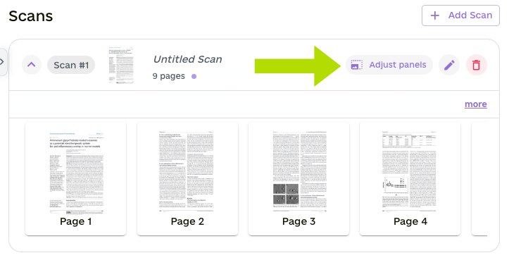
Manual panel adjustment
This feature allows you to manually adjust, update, or delete image panels scanned for integrity issues. While our algorithm automatically extracts figures and sub-image panels to check for integrity issues, you can use this feature for transparency—to see what will be scanned and to adjust the image areas if needed.
You can open the panel editor on the scan page after selecting a paper. Then, our software will automatically precompute figure and sub-image boxes that you can adjust.
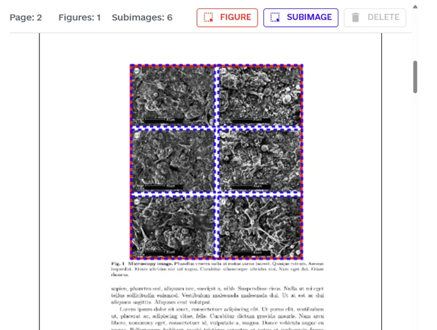
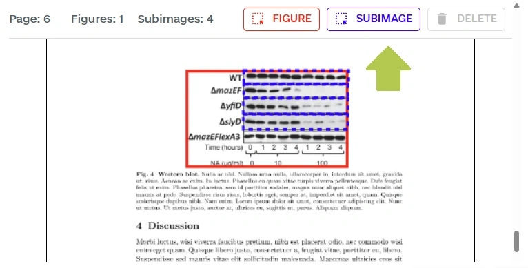
Sub-images: refer to individual panels or sections within a composite research figure. Our software analyzes both entire figures and sub-images for potential integrity issues. Common sub-image types are, for example, western blots, FACS plots, or microscopy image panels. Our algorithm compares sub-images against other sub-images in your paper and against our image database to detect reuse across papers.
Figures: refer to actual figures within your paper, typically labeled with a number and caption. Our algorithm compares figures against our figures in our image database to detect reuse across papers.
You can open the manual panel adjustment on the scan page after selecting a paper.
In this example, one figure (red box) and six subimages (blue boxes) have been correctly labeled. Therefore, no manual adjustment is necessary.
In this example, the automatic extraction missed one subimage — the western blot in the last row. You can draw a new subimage using the button in the top bar.
Important Tabs
PDF report and Sharing your Results
You can generate and download a PDF report and/or share your results after reviewing your results. With the eye icon right next to each finding, you can select what should or shouldn’t be in the report. You can write comments for each finding that will also be displayed in the report.
Confidence Levels
The confidence score can be adjusted from 0% to 100% and reflects the likelihood of a duplicate being a research integrity issue. Scores are computed by our AI models based on the duplicate’s context:
0–32% (Low): Likely a non-problematic duplicate and hidden by default. 33–65% (Fair): Likely a problematic case. 66–100% (High): Highly likely a problematic case.
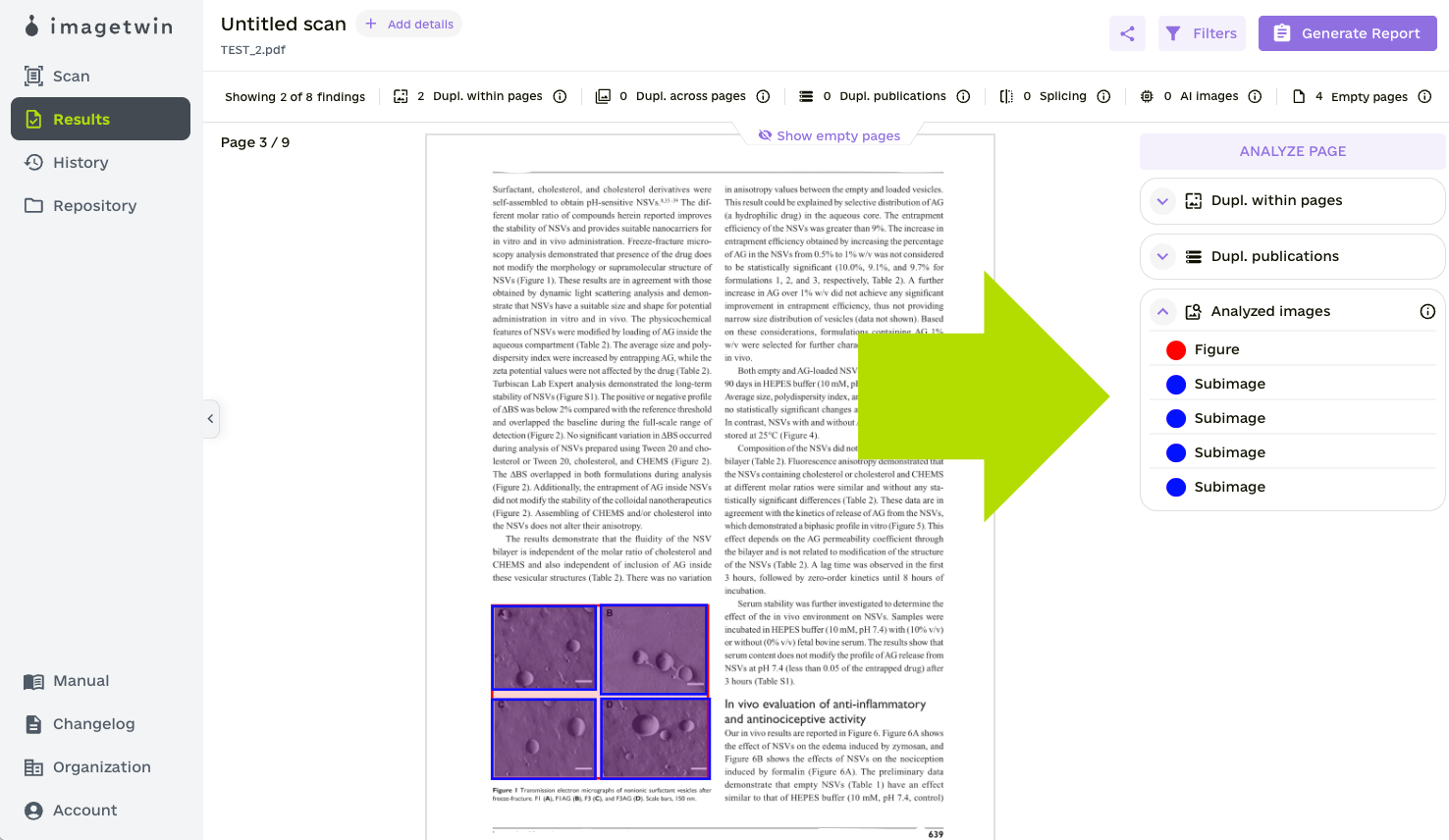
Analyzed Images
You can display the images that were automatically extracted and analyzed by Imagetwin. Use the button shown in the illustration below to display all subimages and figures scanned for integrity issues. We implemented this feature to foster transparency, allowing you to identify which images were analyzed and which were not.
Here is how Imagetwin analyzes sub-images and figures: Extracted subimages (e.g., western blot, microscopy, or FACS panels) are checked individually (e.g., for splicing detection), compared with each other, and matched against our image database to detect reuse across publications. Figures are checked against our image database to detect the reuse of entire figures across articles.
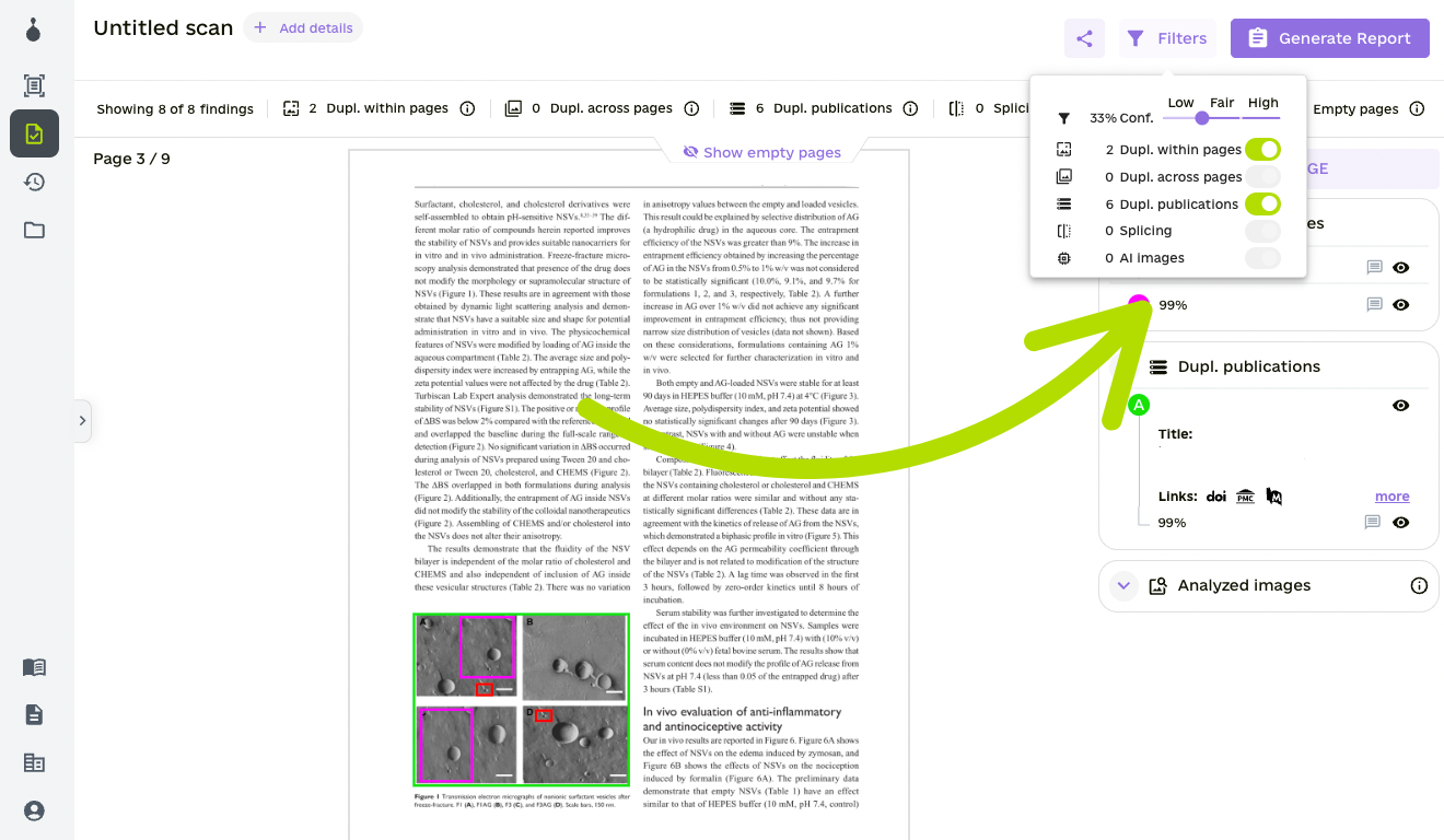
Filters
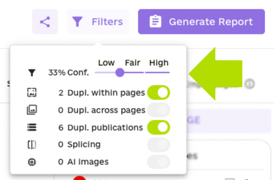
On the top right, you will find the filters.
Duplicates within pages: Filter duplicates that have been detected within your paper/document.
Duplicates across pages: Filter duplicates that have been detected across your paper/document.
Duplicates publications: Filter duplicates that have been detected across publications.
Splicing: Filter splicing detections in gel band images (western blots)
AI Images: Filter AI-generated image detection
In addition to these filters, you can also hide/show duplicates for each external paper where duplicates across publications were detected.
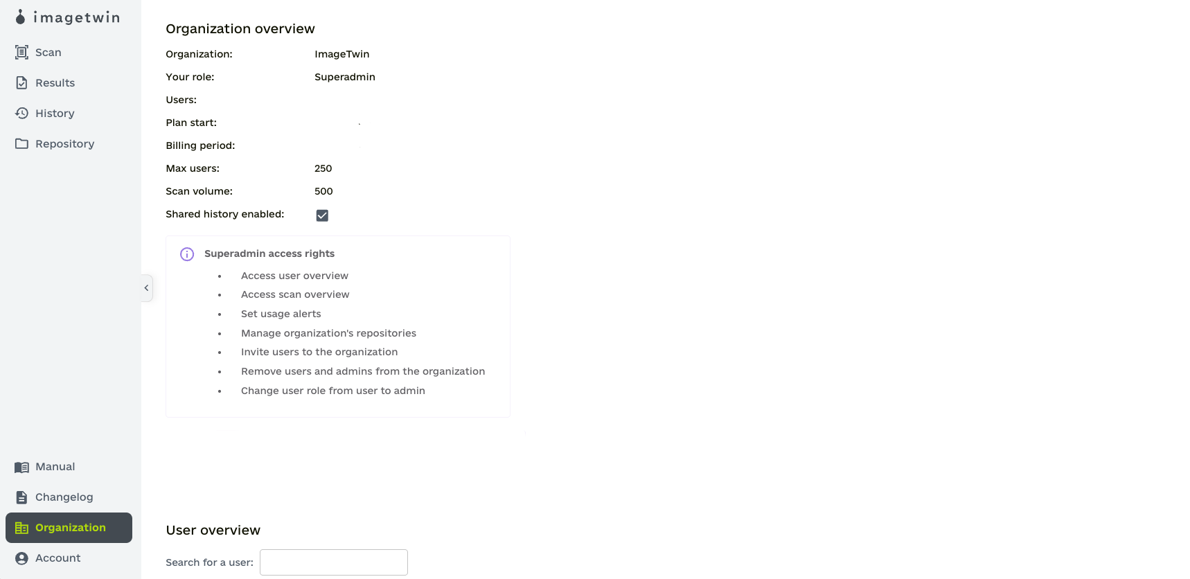
Organization Management
In case you are part of an organization, Admins and Superadmins will have access to an Organization Overview. You can open the overview on the bottom left side under your account.
As Admin, you can:
Access user overview
Access scan overview
Set usage alerts
Invite users to the organization
Remove users from the organization
Change user role from user to admin (as a Superadmin only)